Glaucoma

Glaucoma is a disease in which elevation of eye pressure causes damage to the optic nerve. The optic nerve contains over 1 million neurons which originate in the retina and transmit vision to the brain. If a sufficient number of these neurons are damaged, loss of vision will occur. The side vision is affected initially. In advanced cases, central vision may be permanently lost.
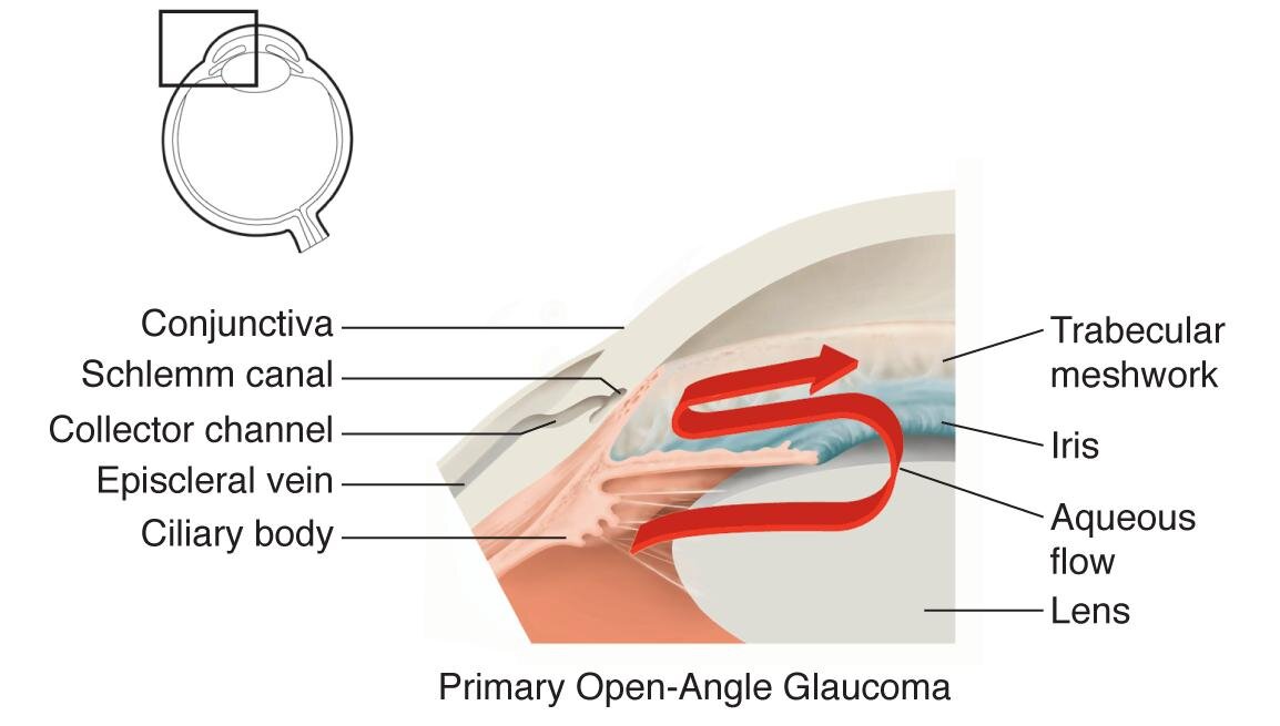
In a normal eye, clear fluid (aqueous) is produced inside the eye by the ciliary body, then drained internally through the open space at the junction of the iris and cornea called the angle. The angle contains the trabecular meshwork and Schlemm’s canal, structures in which the aqueous fluid drains through. Glaucoma occurs when the drainage of fluid is impeded or slowed, and pressure rises. Glaucoma is more common in older patients, African Americans, and those with a family history of the condition. It sometimes occurs in people with normal eye pressure (normal pressure glaucoma).
Types of Glaucoma
Primary
Open-angle glaucoma (OAG) is the most common type of glaucoma, and occurs when aqueous drainage is mildly decreased. The eye pressure slowly increases, but the majority of patients remain asymptomatic. Loss of vision and optic nerve damage are detected during eye examination by the eye doctor.
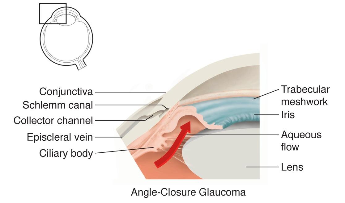
Closed-angle glaucoma, or narrow-angle glaucoma (NAG), occurs when the drainage angle becomes anatomically closed. Eye pressure rapidly increases and causes severe pain and vision loss. Acute angle closure glaucoma is an emergency.
Secondary
Neovascular glaucoma (NVG) occurs when retinovascular disease induces abnormal blood vessel growth on the iris. This seals the angle and blocks aqueous drainage. NVG causes severe pressure, pain, and vision loss.
Steroid medications can cause elevation of eye pressure and subsequently lead to steroid-induced glaucoma.
Uveitic glaucoma is caused by an inflammatory condition of the eye called uveitis, which leads to clogging and scarring of the angle. Steroids are used to treat uveitis and may also contribute to eye pressure elevation.
Angle-recession glaucoma is the result of blunt trauma to the eye causing damage to the trabecular meshwork, leading to reduced aqueous drainage.
Pigmentary glaucoma is a condition in which pigment granules break free from the iris and ciliary epithelium, and obstruct the trabecular meshwork.
Evaluation
A careful review of the patient’s history, including family history, is performed to search for risk factors for glaucoma
The physical exam evaluates the eyes for evidence of glaucoma. Visual acuity and eye pressure is measured. A careful exam of anterior structures including the cornea, anterior chamber, iris, lens, and angle is performed.
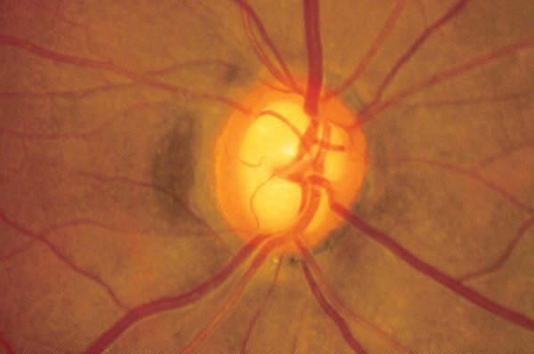
Posterior exam determines if any damage to the optic nerve is apparent. This presents as nerve cupping and thinning, usually of the inferior and/or superior nerve.
Fundus photographs are taken to document the appearance of the nerve.
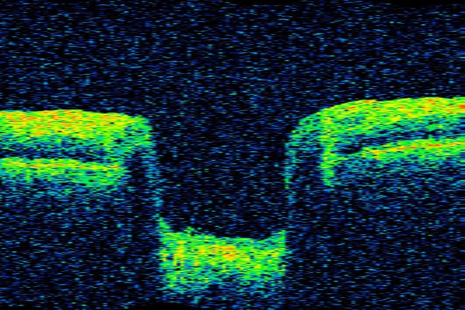
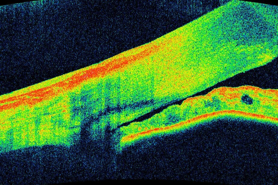
Optical coherence tomography (OCT) is a scan which measures the neurons of the optic nerve with great accuracy. It is able to detect glaucoma at its earliest stages, before damage to the vision has occurred. OCT can also visualize the front structures of the eye, including the angle.
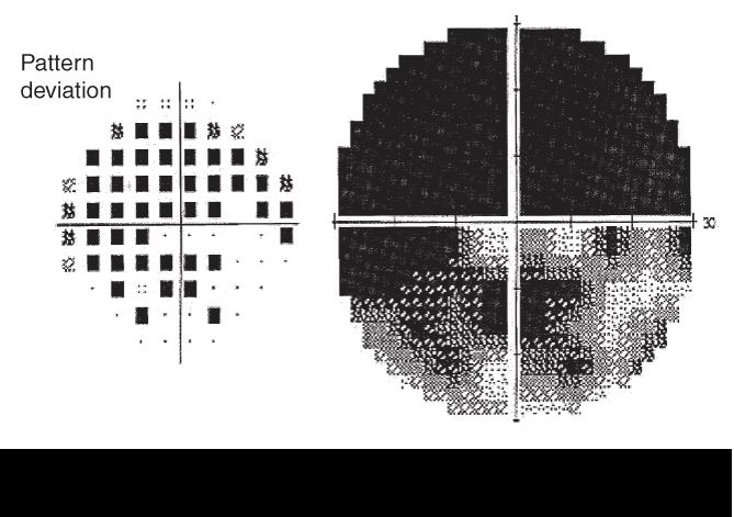
Visual field testing is performed to assess the overall visual function of the eye. Special attention is directed to the peripheral vision to detect the earliest damage to the patient’s vision.
From the Expert…
Glaucoma is a disease in which neurons, traveling from the retina to the brain’s visual cortex via the optic nerve, are damaged. The neurons die and the vision they provide is lost.
Glaucoma is a disease of progression, as neurons cannot be rejuvenated. Therefor early detection is critical for a successful outcome. Careful monitoring of the patient’s status guides therapy, is the standard of care, and can usually allow prevention of vision loss.
Additional Resources
American Academy of Ophthalmology Glaucoma Panel. Preferred Practice Pattern®Guidelines. Primary Open-Angle Glaucoma. San Francisco, CA: American Academy of Ophthalmology; 2015. Available at: www.aao.org/ppp.
American Academy of Ophthalmology Glaucoma Panel. Preferred Practice Pattern®Guidelines. Primary Angle Closure. San Francisco, CA: American Academy of Ophthalmology; 2015. Available at: www.aao.org/ppp.
Glaucoma. American Academy of Ophthalmology. EyeWiki. https://eyewiki.aao.org/Category%3AGlaucoma. Accessed August 29, 2019.
Summary Benchmarks for Preferred Practice Pattern® Guidelines. Glaucoma. American Academy of Ophthalmology; 2018.