Posterior Vitreous Detachment & Retinal Tear
Posterior Vitreous Detachment
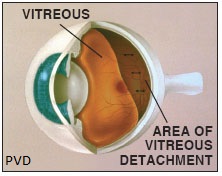
Posterior Vitreous Detachment (PVD) is a condition that occurs when the vitreous gel separates from the retina. This is a normal part of the aging process.
Retinal Tear
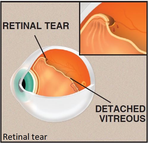
In some cases, when a PVD occurs, the vitreous separating from the retina can pull on an area of the retina, creating a tear. Retinal tears can occur in anyone, but are more common in the elderly, in nearsighted individuals, or those with abnormally strong attachments between the retina and vitreous. Some retinal tears are due to trauma.
SYMPTOMS
Most people who experience a PVD are suddenly aware of new floaters caused by the now-contracted and separated vitreous floating visibly in front of the retina. In addition to new floaters, some patients with PVD observe flashing lights that occur due to the vitreous pulling on the retina, typically described as sparks or split-second, arc-like flashes located in the peripheral vision.
EVALUATION
A complete ocular examination, including a dilated fundus examination, is indicated if a PVD is suspected.
Fundus photography is useful to search for and document the presence of the PVD and any tears when present.
Ultrasonography may be performed to evaluate the status of the vitreous. It is also useful to determine if the retina is detached, especially if the view is obscured by vitreous hemorrhage.
Optical Coherence Tomography (OCT) can be performed to help determine if the vitreous has separated from the retina. It may also be useful to evaluate for the presence of subretinal fluid.
TREATMENT
PVDs are usually observed. If the floaters significantly impact the quality of the patient’s vision, they can be removed with a surgical technique called pars plana vitrectomy. It may be prudent to wait several months before proceeding to vitrectomy to see if the patient is able to adapt to the floaters.
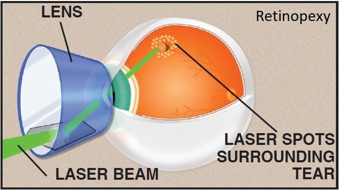
Retinal tears usually are treated with laser. Retinal laser causes an adhesion between the retina and the eye wall in a circular configuration around the retinal tear. This bond can usually keep the tear from progressing to a retinal detachment.
COMPLICATIONS
The primary goal in managing patients with PVD is to detect and treat retinal tears. If left untreated, many tears lead to retinal detachment, in which the retina separates from the back wall of the eye.
Complications of laser treatment are uncommon. It can cause bleeding at the point of laser contact with the retina.
Importantly, even with thorough evaluation and appropriate treatment, the retina may still detach. This is either from holes that weren’t treated, fluid tracking through the laser, or new holes which may form.
From the Expert…
One of the most frequent conditions ophthalmologists encounter is posterior vitreous detachment. While the condition is relatively common, complications such as vitreous hemorrhage, retinal tear, or retinal detachment are not. As the symptoms of PVD, retinal tear, and retinal detachment are similar, a complete examination including dilated fundoscopy, and sometimes retinal imaging or ultrasonography, should be performed in a timely manner.
In my experience, approximately 10% of patients with PVDs will have a retinal tear.
Retinal tears, when diagnosed early, are usually treated in the office with laser or cryotherapy. For providers who lack the capability to treat the patient themselves, timely referral is indicated. With appropriate care, progression of the disease and subsequent vision loss can usually be avoided. That is why the standard of care is for prompt evaluation and treatment when indicated.
Additional Resources
American Academy of Ophthalmology Retina/Vitreous Panel. Preferred Practice Pattern®Guidelines. Posterior Vitreous Detachment, Retinal Breaks, and Lattice Degeneration. San Francisco, CA: American Academy of Ophthalmology; 2014. Available at: www.aao.org/ppp.
Tabernero, S. Posterior vitreous detachment. American Academy of Ophthalmology. EyeWiki. https://eyewiki.aao.org/Posterior_vitreous_detachment. Accessed June 21, 2019.
Retina Health Series. Posterior Vitreous Detachment. The Foundation of the American Society of Retina Specialists. https://www.asrs.org/content/documents/fact_sheet_1_posterior_vitreous_detachment_new.pdf. Accessed June 21, 2019.
Retina Health Series. Retinal Tears. The Foundation of the American Society of Retina Specialists. https://www.asrs.org/content/documents/fact_sheet_23_retinal_tears_new.pdf. Accessed June 21, 2019.
Summary Benchmarks for Preferred Practice Pattern® Guidelines. Posterior Vitreous Detachment, Retinal Breaks, and Lattice Degeneration. American Academy of Ophthalmology; 2018.